



B-hACP3 PC-3

NA • 322552

B-hACP3 PC-3

Catalog Number: 322552
Strain Name: NA
NCBI gene ID: 55 (Human)
Aliases: ACPP; 5'-NT; ACP-3; TM-PAP
---
라이선스 옵션 제공 가능
B-hACP3 PC-3

이 페이지에서

  • Description
  • Phenotypic analysis
  • Tumorigenicity

포스터

모두 보기

    출판물

      Description
      • Origin: The PC-3 cell line is an androgen-independent model derived from a bone metastasis of a high-grade prostatic adenocarcinoma in a 62-year-old male. It serves as a critical in vitro tool for advancing research in prostate cancer biology, particularly for investigating mechanisms of tumor progression, metastasis, and the development of therapeutic resistance.
      • Background Information: Acid phosphatase 3 (ACP3), also known as prostatic acid phosphatase (PAP), as an ecto-phosphatase that hydrolyzes extracellular nucleotides and phosphoproteins, influencing cell growth, migration, and invasive properties. ACP3 exhibits high tumor specificity, with high expression in prostate cancer tissues but minimal expression in non-cancerous tissues, making it a therapeutic target for prostate cancer.
      • Gene targeting strategy: The exogenous promoter, human ACP3 coding sequences were inserted to AAVS1 locus in B-hACP3 PC-3.
      • Tumorigenicity: Confirmed in B-NDG mice.
      • Application: The B-hACP3 PC-3 tumor models can be used for preclinical evaluation of monoclonal antibody drugs targeting human ACP3.
      ACP3 Protein Expression Analysis
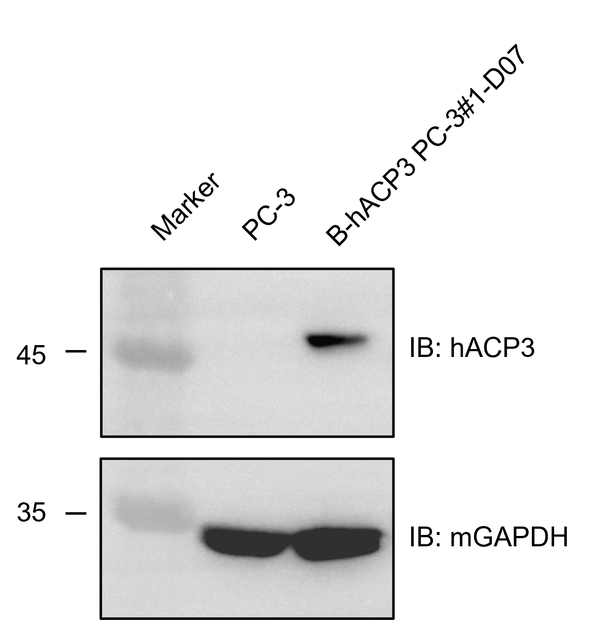
      • Human ACP3 was detected in B-hACP3 PC-3 but not in wild-type PC-3 cells.

      ACP3 expression analysis in B-hACP3 PC-3 by western blot. Single cell suspensions from wild-type PC-3 and B-hACP3 PC-3 cultures were stained with anti-hACP3 antibody (CST, 12861S). Human ACP3 was detected in the B-hACP3 PC-3 cells but not in wild-type PC-3 cells. The 1-D07 clone of B-hACP3 PC-3 cells was used for in vivo experiments.


      Tumor Growth Curve & Body Weight Changes

      Subcutaneous homograft tumor growth of B-hACP3 PC-3 cells. B-hACP3 PC-3 cells (5x10⁶) and wild-type PC-3 cells (5x10⁶) were subcutaneously implanted into B-NDG mice (male, 10-week-old, n=6). Tumor volume and body weight were measured twice a week. (A) Average tumor volume ± SEM. (B) Body weight (Mean± SEM). Volume was expressed in mm3 using the formula: V=0.5 X long diameter X short diameter2. As shown in panel A, B-hACP3 PC-3 cells were able to establish tumors in vivo and can be used for efficacy studies.
      ACP3 Protein Expression Analysis of Tumor Tissue
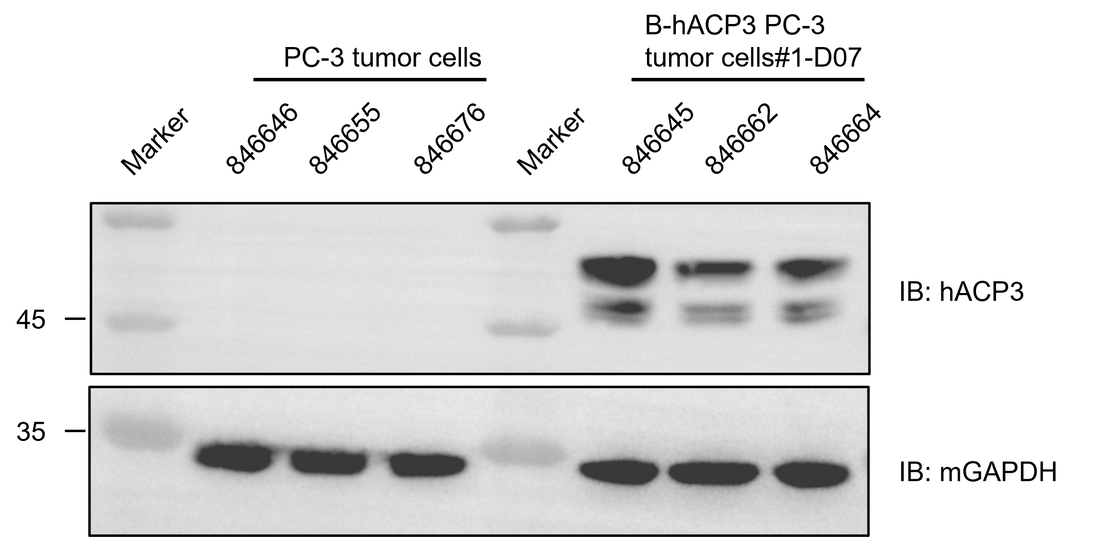
      • Human ACP3 was expressed in B-hACP3 PC-3 tumor cells, but not in PC-3 tumor cells.

      Human ACP3 expression evaluated in B-hACP3 PC-3 tumor cells by western blot. B-hACP3 PC-3 cells were subcutaneously transplanted into B-NDG mice (Male, 8-week-old, n=6). At the end of the experiment, tumor cells were harvested and assessed with anti-human ACP3 antibody (CST, 12861S).
