
• 322270

The mouse Cdh17 gene was replaced by human CDH17 coding sequence in B-hCDH17 B16-F10 cells. Human CDH17 is highly expressed on the surface of B-hCDH17 B16-F10 cells.

Gene targeting strategy for B-hCDH17 B16-F10 cells. The exogenous promoter and human CDH17 coding sequence was inserted to replace part of murine exon 3.

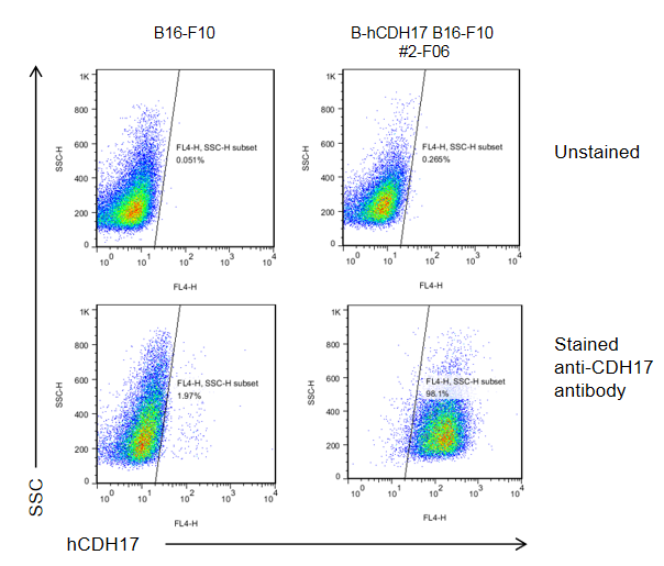
CDH17 expression analysis in B-hCDH17 B16-F10 cells by flow cytometry. Single cell suspensions from wild-type B16-F10 and B-hCDH17 B16-F10 cultures were stained with anti-CDH17 antibody (provided by a corporate partner). Human CDH17 was detected on the surface of B-hCDH17 B16-F10 cells but not wild-type B16-F10 cells.

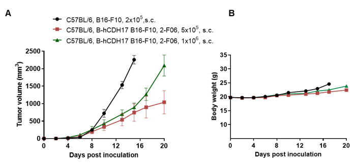
Subcutaneous homograft tumor growth of B-hCDH17 B16-F10 cells. B-hCDH17 B16-F10 cells and wild-type B16-F10 cells were subcutaneously implanted into C57BL/6 mice (female, 9-week-old, n=8). Tumor volume and body weight were measured twice a week. (A) Average tumor volume ± SEM. (B) Body weight (Mean± SEM). Volume was expressed in mm3 using the formula: V=0.5 X long diameter X short diameter2. As shown in panel A, B-hCDH17 B16-F10 cells were able to establish tumors in vivo and can be used for efficacy studies.
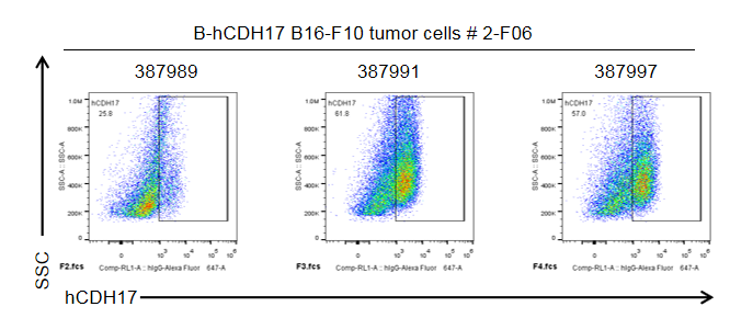
Tumor cells were harvested at the end point of the experiment and assessed for human CDH17 expression by flow cytometry. As shown in the panel, human CDH17 was expressed on the surface of tumor cells (anti-human CDH17 antibody was provided by a corporate partner).
Note. B16-F10 tumor cells harvested at the end point of the experiment are easily dead, which may influence the protein expression analysis.
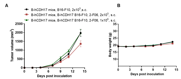
Subcutaneous homograft tumor growth of B-hCDH17 B16-F10 cells. B-hCDH17 B16-F10 cells and wild-type B16-F10 cells were subcutaneously implanted into B-hCDH17 mice (female, 9-week-old, n=6). Tumor volume and body weight were measured twice a week. (A) Average tumor volume ± SEM. (B) Body weight (Mean± SEM). Volume was expressed in mm3 using the formula: V=0.5 × long diameter × short diameter2. As shown in panel A, B-hCDH17 B16-F10 cells were able to establish tumors in vivo and can be used for efficacy studies.
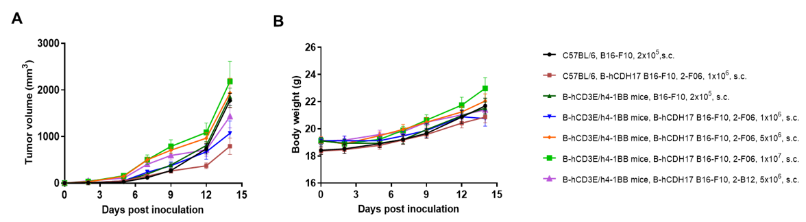
Subcutaneous homograft tumor growth of B-hCDH17 B16-F10 cells. B-hCDH17 B16-F10 cells and wild-type B16-F10 cells were subcutaneously implanted into C57BL/6 mice or B-hCD3E/h4-1BB mice (female, 7 to 8-week-old, n=8) respectively. Tumor volume and body weight were measured twice a week. (A) Average tumor volume ± SEM. (B) Body weight (Mean± SEM). Volume was expressed in mm3 using the formula: V=0.5 × long diameter × short diameter2. As shown in panel A, B-hCDH17 B16-F10 cells were able to establish tumors in vivo.
