

B-hEPCAM mice(C)
BALB/cCrSlcNifdc-Epcamtm1(EPCAM)Bcgen/Bcgen • 114390

B-hEPCAM mice(C)

Catalog Number
114390
Strain Name
BALB/cCrSlcNifdc-Epcamtm1(EPCAM)Bcgen/Bcgen
Strain Background
BALB/cCrSlcNifdc
NCBI gene ID
4072 (Human)
Aliases
Ber-Ep4, BerEp4, DIAR5, EGP-2, EGP314, EGP40, ESA, HNPCC8, KS1/4, KSA, LYNCH8, M4S1, MIC18, MK-1, MOC-31, TACSTD1, TROP1

이 페이지에서

  • Description
  • Phenotypic analysis

포스터

모두 보기

    출판물

      Description

       EPCAM: A key adhesion molecule in tumor malignancy and its therapeutic intervention

      • Gene Information: EPCAM is a transmembrane glycoprotein featuring both extracellular (EpEx) and intracellular (EpICD) domains. Cleavage of EPCAM releases EpICD, which translocates to the nucleus as a transcriptional co-factor.
      • Protein Expression: EPCAM is expressed in various human epithelial tissues, cancerous tissues, progenitor cells, and stem cells. It is widely overexpressed in a variety of solid tumors and tumor-initiating cells (TICs), playing a vital role in fueling cancer progression, particularly in colorectal cancer (CRC).
      • Signaling Pathway: EpICD interacts with Wnt receptor promoters (FZD6 and LRP5/6) to upregulate their transcription and activate Wnt signaling. Additionally, activation of Wnt kinases (GSK3β and CK1) in the β-catenin destruction complex induces γ-secretase activity to enhance EpICD shedding, forming a positive feedback loop.
      • Therapeutic Inhibition: By blocking EPCAM-mediated Wnt receptor upregulation and signaling, thereby inhibiting tumor growth and prolonging survival in CRC models.
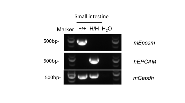
      Human EPCAM mRNA Expression Analysis in EPCAM Humanized Mice (BALB/c)

      Human EPCAM was detectable in EPCAM Humanized Mice by RT-PCR and sequencing. Small intestine tissues were isolated from wild-type BALB/cCrSlcNifdc mice (+/+) and homozygous B-hEPCAM mice(C) (H/H). Primers were designed to detect mouse and human EPCAM. Sequencing of the PCR products confirmed that the amplified sequences were consistent with database reference sequences.
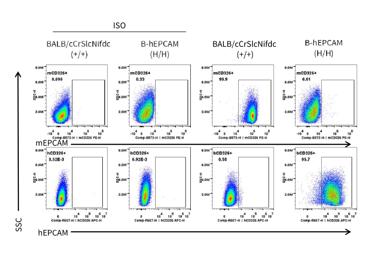
      Human EPCAM Protein Expression Analysis in Small Intestine of EPCAM Humanized Mice (BALB/c)

      Strain-specific EPCAM expression analysis in wild-type BALB/cCrSlcNifdc mice and homozygous EPCAM humanized mice. Small intestine cells were collected from wild-type BALB/cCrSlcNifdc mice and homozygous B-hEPCAM mice (C). EPCAM expression was analyzed by flow cytometry using anti-human EPCAM antibody (Biolegend, 369809) and anti-mouse EPCAM antibody (Biolegend, 118205).
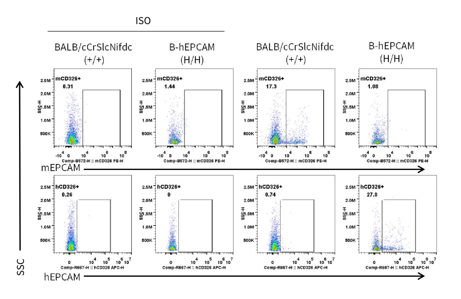
      Human EPCAM Protein Expression in Lung of EPCAM Humanized Mice (BALB/c)

      Strain specific  EPCAM expression analysis in wild-type BALB/cCrSlcNifdc and homozygous  EPCAM humanized mice by flow cytometry. Lung cells were collected from wild-type BALB/cCrSlcNifdc mice and homozygous B-hEPCAM mice(C). EPCAM expression was analyzed by flow cytometry using anti-human EPCAM antibody (Biolegend, 369809) and anti-mouse EPCAM antibody (Biolegend, 118205).
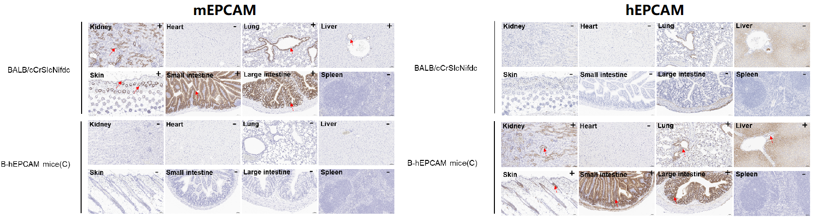
      Protein Expression Profiles of EPCAM in EPCAM Humanized Mice (BALB/c)
      • Mouse EPCAM was detectable in BALB/cCrSlcNifdc. Human EPCAM was detectable in EPCAM humanized mice.
      • Mouse and human EPCAM were both detected in kidney, lung, liver, skin, small intestine and large intestine, but not in heart and spleen. 

      Immunohistochemical (IHC) analysis of EPCAM expression in EPCAM humanized mice. The kidney, heart, lung, liver, skin, small intestine, large intestine and spleen were collected from wild-type BALB/cCrSlcNifdc mice and B-hEPCAM mice(C) (female, 6-week-old), analyzed by IHC with anti-mEPCAM antibody (abcam, ab213501) and anti-hEPCAM antibody (abcam, ab223582). The arrow   indicates tissue cells with positive EPCAM staining (brown). “+” indicates that the tissue is positive, and “-” indicates that the tissue is negative.

      * When publishing results obtained using this animal model, please acknowledge the source as follows: The animal model [B-hEPCAM mice(C)] (Cat# 114390) was purchased from Biocytogen.